Digital Radiography in Dentistry
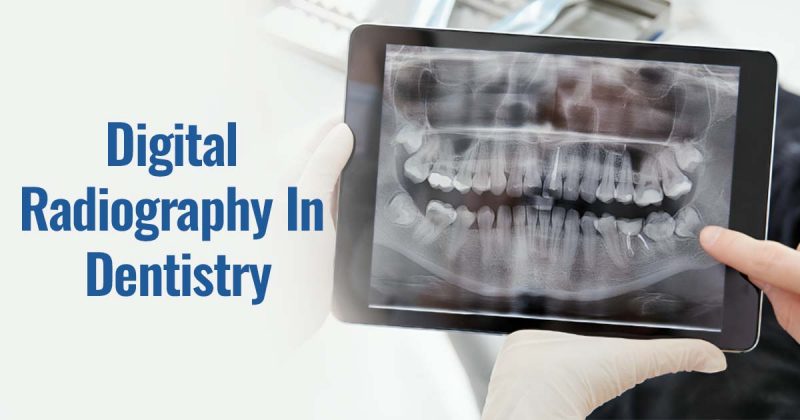
Digital Radiography in Dentistry – The dynamic and modern era of dental imaging is characterized by digital radiography, which is one of the greatest advances in the industry. No longer do we have to put up with heavy films and dark developing rooms; rather we are now using a modern device for taking detailed images of the teeth instantly. In this blog, we will look into the meaning of digital radiography, examine the different types of digital dental radiographs, and learn why this new methodology is rapidly overtaking the conventional ones. Let’s explore the scope of dental imaging in the years to come!
What is digital radiography in dentistry?
Digital radiography, usually called digital X-rays, is a superior imaging technique used in dentistry where electronic sensing devices are used instead of traditional radiographic films. Such images may include those of the teeth and other maxillofacial structures. Its primary functions are related to the prevention, diagnosis, and treatment of dental diseases. This technology enables the immediate acquisition and display of high-quality images, which can be enhanced for better diagnosis and treatment planning.
Types of Digital Dental Radiographs:
Digital dental radiographs can be of two types: intraoral or extraoral, depending on how they are being radiographed, either internally or externally.
Intraoral Radiographs:
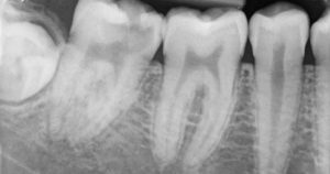
1. Periapical X-rays- Here, the X-ray image taken with the help of a sensor helps in viewing the crown up to the root region including the surrounding bone at the apex in the upper or the lower jaw whichever is targeted. These X-rays help in detecting endodontic and periodontal lesions, tooth decay, bone loss, etc.
Extraoral Radiographs:
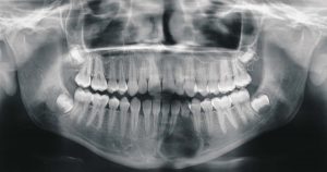
1. Panoramic X-rays – More specifically known as orthopantomogram or OPG, this is a device that rotates around the head of the patient to provide an image of the teeth in relation to the jaws. The orthopantomogram shows both the upper and lower jaws and the entire bone structure in one picture. OPGs often demonstrate the position of the developing teeth; more importantly, they also assist in identifying tumors, impacted third molars, or any other irregularity occurring in the jaws.
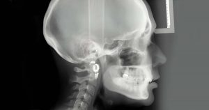
2. Cephalometric X-rays – This specific view depicts the profile of the head specifically highlighting the coordinates of dental and skeletal structures along with the soft-tissue profiles. This is mainly utilized by orthodontists for assessing the malalignment of teeth, planning their respective treatments, and verifying the post-treatment results too.
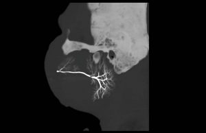
3. Sialography – This is a specialized imaging technique proposed only for assessing the morphology and functionality of the parotid and submandibular glands. The principal indication for sialography is chronic inflammatory conditions especially where there is obstruction as in Sjogren’s syndrome. The technique involves infusion of the gland ductal system with an iodinated contrast agent, and then imaging the gland with projection imaging, fluoroscopy, or cone beam computed tomography (CBCT).
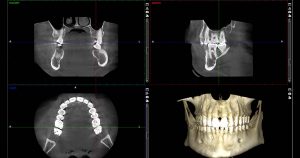
4. Cone beam computed tomography – It acquires data volumetrically providing three–dimensional radiographic images for the assessment of the dental and maxillofacial complex, facilitating dental diagnosis. Used to detect potential extra canals before endodontic treatment, pre-surgical planning for an implant, third molar positionings, benign calcifications, cysts, and tumors in the jaw.
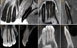
5. Magnetic Resonance Imaging- MRI is non-invasive and uses non-ionizing radiation. It makes images with excellent soft-tissue resolution in any imaging plane without reorienting the patient. The primary limitations of MRI relate to the prolonged periods required for scanning and the cost involved. It’s useful in assessing the oral cavity for jaw tumors, and any vascular anomaly, and in early assessment of jaw osteomyelitis when there is a suspicion.
Why are traditional radiographs getting out of trend and digital radiographs gaining popularity?
1. Instant results: Digital dental radiographs are processed instantly whenever the targeted area is exposed. This helps in quicker diagnosis and treatment procedures for the dentist thus giving him/her more time to look after other patients in the daily practice and at the same time providing the patients with short-period dental appointments. Thus, saving time and discomfort for both of them.
2. Good image quality: Digital radiographs provide images of better quality and can be electronically edited for better image definition. They can be magnified to check for any minute changes, and contrast and sharpness can also be adjusted in the targeted structures for a better view. These images can further be flipped or colorized as per the need.
3. Less radiation exposure: The radiation exposure in digital radiographs is about 80% less than the radiation used in traditional x-rays as the sensors used in this are more sensitive. Hence, reducing patients’ radiation exposure time and dosage.
4. Micro–storage: In digital radiography, patient’s data is stored electronically thus reducing the storage space. These stored documents are easy to access and can be shared with the patients, consultant dentists, and insurance companies for claims, way more easily. This data is less likely to be lost or misplaced when compared to x-ray films. Additionally, the data is easily sorted according to separate files for each patient which otherwise could have been a tedious task with the previously used x-ray films.
5. Environment friendly: Digital radiographs are eco-friendly with no usage of chemicals for processing and disposal of hazardous materials like lead foils, leading to a decrease in the landfill. Digital radiographs also eliminate the need for a dark room which was once needed for processing an X-ray film thus, reducing the need for more clinical space.
Read Also: How Advances in Oral Radiology and Medicine Are Changing Dentistry
Disadvantages of digital radiography:
1. High cost – Digital radiography costs much higher initially and requires a good amount of investment in the computer, software, hardware, servers, and wifi connections which may be a problem for dentists with small-scale dental clinic setups.
2. Dependency on technology: The technology used in digital radiography requires frequent updates which when not updated can hinder patients’ treatment procedures or delay them.
3. Over-reliance – The ease of getting a digital image leads to over-reliance on them for diagnosis which can end up a patient being overly exposed to radiation.
4. Sensor fragility- The sensors used in digital radiography are highly fragile and require care while handling to prevent any damage to them.
5. Infection control – Infection control is not as good as the sensors and other machinery cannot be completely sterilized thus increasing the chances of cross-contamination.
Conclusion
To sum up, digital X-ray has become a game changer in the field of dentistry, providing numerous benefits over conventional ways. Given the features- instant imaging, better quality, less radiation, and green practices, of course, dentists are making the change. There are a few obstacles to overcome, but the pluses are greater than the minuses. In the long run, it presents a bright future for the professionals and in turn for the patients too; it encourages the use of digital radiography in patient care as well as the development of improved dental diagnostic techniques. For all your digital radiography needs, DentalKart offers a comprehensive selection of dental equipment online. Explore our page to discover everything you need with just a click—because the future of dental imaging is bright, and digital radiography is leading the way!

No Comment