Mastering Suturing Techniques and Choosing the Right Materials in Dentistry

Oral surgery is more than just making incisions and extracting teeth — it’s about ensuring that every wound heals smoothly, comfortably, and without complications. One of the key elements in achieving this is the proper selection and application of sutures. The right suture material, combined with the correct suturing technique, plays a critical role in promoting tissue healing, minimizing discomfort, and preventing postoperative infections.
From simple tooth extractions and dental implant placements to more complex procedures like flap surgeries, bone grafting, soft tissue grafting, corrective jaw surgeries, and facial trauma repair — suturing is a fundamental skill that underpins successful outcomes in nearly all aspects of oral surgery. Each procedure demands a tailored approach, depending on the location, tissue type, and desired healing response.
Whether it’s stabilizing tissue after a sinus lift, securing a flap in TMJ surgery, or closing incisions after the removal of cysts, tumors, or impacted teeth, the choice of suture can make a significant difference.
In this blog, we’ll dive into the essentials of dental sutures — from the types of suture materials to the principles and techniques of suturing commonly used in oral surgical procedures. Let’s stitch together the knowledge needed for better healing and better smiles!
What is a Suture?
A suture is a stitch, or a series of stitches made to secure the apposition of the edges of a surgical/ traumatic wound.
A suture material is an artificial fibre used to stitch the traumatised tissue together until they hold sufficiently well by themselves natural fibre(collagen), which is synthesised and woven into a strongest scar.
Classification of Suturing Materials:
Suturing materials can be of following types depending on various criteria:
- According to absorbability: Absorbable or Non-absorbable
- According to sources: Natural or Synthetic
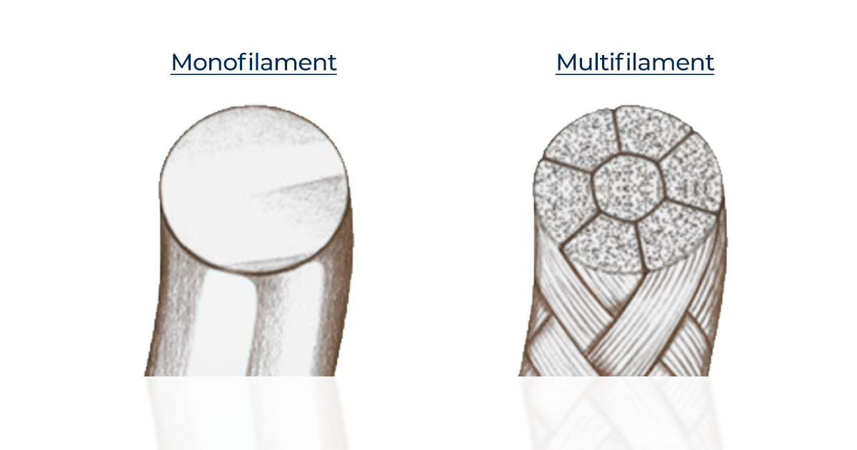
- According to structure: Monofilament or Multifilament
- According to coating: Coated or Uncoated, Dyed or Undyed
- According to tissue reaction: Reactive or Non-reactive
Absorbable Suture:
These sutures are popular in periodontal and implant surgeries, as they cause less postoperative inflammation, have less absorbable time, and provide more patient comfort. These can be of the following types:
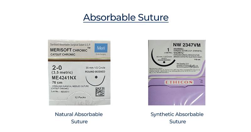
Natural Absorbable Suture:
These are monofilaments of highly purified collagen with mild to moderate tensile strength. One disadvantage is that they can cause a mild inflammatory reaction and can be difficult to handle. They should not be used in high acid environments (like reflux bulimia, esophagitis, Sjogren’s syndrome, radiation therapy, etc.). These can be further divided into two types:
a. Plain catgut, which has a resorption rate of 3 to 5 days.
b. Chromic catgut, which is dyed with a chromium salt solution and has a prolonged resorption rate of 7 to 10 days.
Synthetic Absorbable Suture:
These are braided filaments of polyglycolic acid, which are absorbed by hydrolysis (hydrophobic). They have good tensile strength (resist muscle pull), inhibit bacterial growth, cause only mild tissue reaction, and have a resorption rate of 21 to 28 days.
These can be of the following two types:
a. Dexon: Has a non-toxic coating or is uncoated, violet or undyed.
b. Vicryl: Coated – violet or undyed, stronger than Dexon.
Top Recommendations: Meril Merisoft Chromic #3-0 Catgut Suture, Meril Megasorb #4-0 Polyglycolic Acid Suture, Ethicon Vicryl #1 Absorbable Violet Braided Suture
Non-Absorbable Sutures
These sutures are resistant to hydrolysis and proteolysis and have good tensile strength. Their non-absorbable nature can be discomforting for patients in some cases. These can further be divided into the following two types:
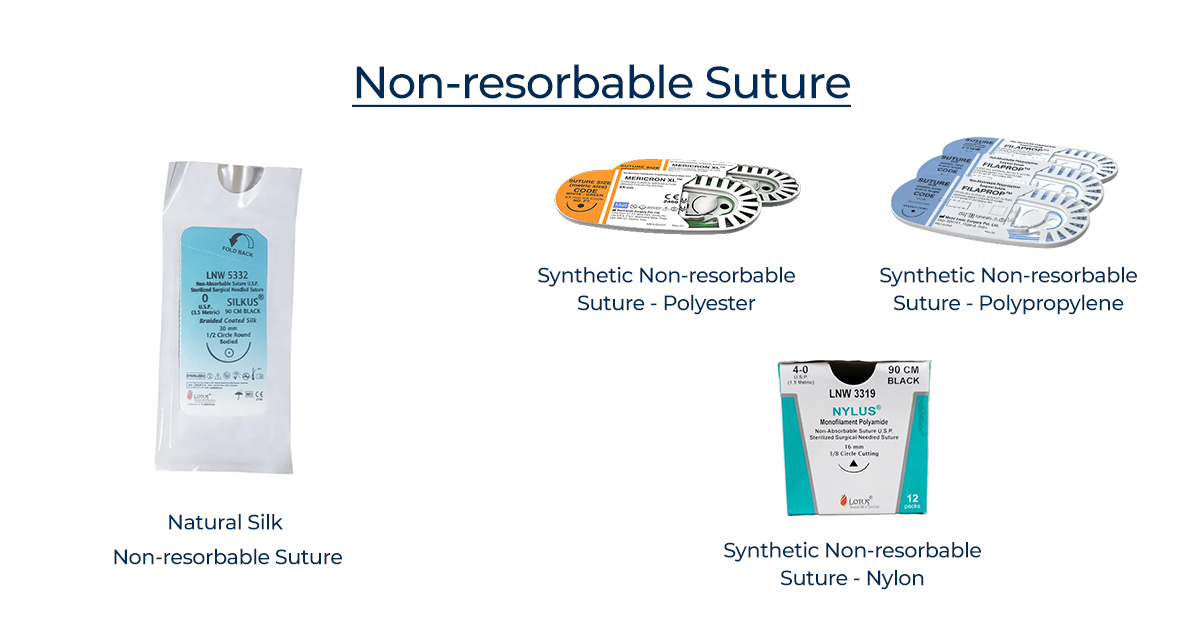
Natural Non-resorbable Suture:
a) Silk: It is a braided, inexpensive, and readily available suture. The braided fibers allow for high elasticity and help the material adhere to itself, maintaining tension within the knot and allowing for secure knots. However, the disadvantage with these sutures is that they are more likely to cause infection, and removal is done one week after placement. The braided arrangement of fibers increases the surface area, which wicks fluid and bacteria into the thread. This causes bacteria to spread into the wound, increasing the likelihood of infection and causing a severe inflammatory reaction.
Silk sutures are thus contraindicated in implant and particulate graft procedures.
b) Cotton/Linen: These are also braided filaments and are easy to handle but are capable of causing severe inflammatory reactions similar to silk sutures. Another disadvantage is that they have poor strength.
Top Recommendations: Lotus Silkus #0 Black Braided Silk Sutures (Pack of 12), Meril Filasilk #5-0 Black Braided Silk Suture
Synthetic Non-resorbable Suture:
a) Nylon: These can be of braided (coated) or non-braided type and have excellent handling qualities along with good knot security. A major advantage is that they have greater tensile strength than silk sutures and cause no inflammatory reaction.
Top Recommendations: Lotus Nylus #4-0 Black Monofilament Polyamide Suture LNW3319 (Pack of 12)
b) Polypropylene: These are monofilament threads with excellent tensile strength and no inflammatory reaction. Their non-absorbable property decreases the likelihood of bacterial growth. These can be difficult to handle and require a surgeon’s knot to prevent them from getting untied.
Top Recommendations: Meril Filaprop #2-0 Polypropylene Suture
c) Polyester: These sutures are braided and can be coated or uncoated. Additionally, these do not cause tissue reaction. They also require a good surgeon’s knot to prevent them from getting untied, as they have a strong material memory.
Top Recommendations: Meril Mericron XL #4-0 Polyester Suture
d) Polytetrafluoroethylene (PTFE) / Gore-Tex: These are monofilament, high tensile strength sutures with excellent handling properties. They cause low tissue reaction like the others but are highly expensive.
Principles of Suturing:
- The needle should be grasped with a needle holder at about three-fourths of its distance from the tip of the needle.
- The needle should never be held at the suture end, as it is the weakest and can lead to bending or breakage of the needle.
- The needle should pierce the tissue perpendicular to its surface, as piercing the tissue obliquely may result in a tear.
- The curvature of the needle should be followed while passing through the tissues to prevent tearing them.
- The suture should be placed equidistant (2–3 mm) from the incision line. The depth of the penetration should be equal on both sides of the line.
- The needle should pass from the mobile tissue to the fixed tissue.
- The needle should pass from the thinner tissue to the thicker tissue.
- Similarly, the needle should pass through the deeper side first and then through the superficial side.
- The distance from the incision point to the needle penetration should be less than the depth to which the needle penetrates into the tissues in order to cause eversion of the wound margin when the suture is tied.
- The suture should just approximate the wound margins and should never be so tight that it causes blanching of the tissues.
- The knot should not be placed over the wound margins.
- Each suture should be placed 3 – 4 mm apart.
- When the length of the tissue on one side of the wound is longer than the other, suturing may result in dog-ear formation, i.e., excessive tissue on one side. To eliminate this, an incision at approximately 30 – 45 degrees to the original incision is directed at the undermined tissue. The extra tissue is pulled over the incision, an appropriate amount is excised, and the wound is closed.
Common Suturing Techniques used in Dentistry:
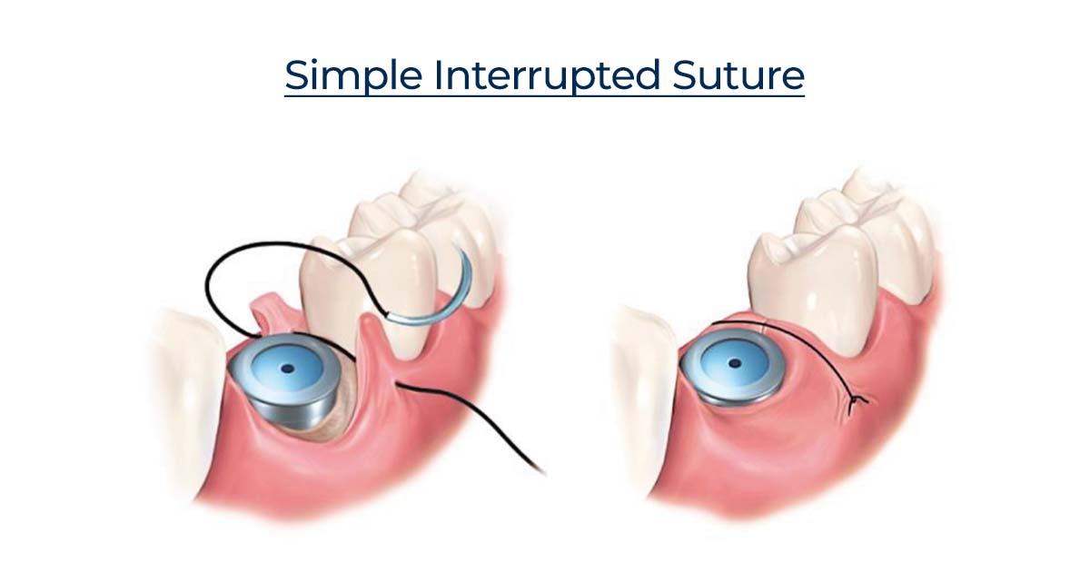
1. Simple Interrupted:
It is one of the most commonly used suturing techniques in dentistry. It requires the needle to first penetrate the buccal gingiva of the operative site, cross the wound, and exit from the lingual side with the needle oriented medially throughout this movement. A loop is created, and the suture thread is tied off at the original entry point. It can be used for the closure of small wounds or can be placed in multiples to close larger wounds.
Advantages:
• Strong and can be placed in areas of stress
• Placed 4–8 mm apart to close large wounds, helping in the distribution of tension
• Each suture is independent, and loosening of one will not cause the other sutures to loosen
• In case of infection or hematoma, removal of some sutures can be done
• Free of interferences between each stitch and easy to clean
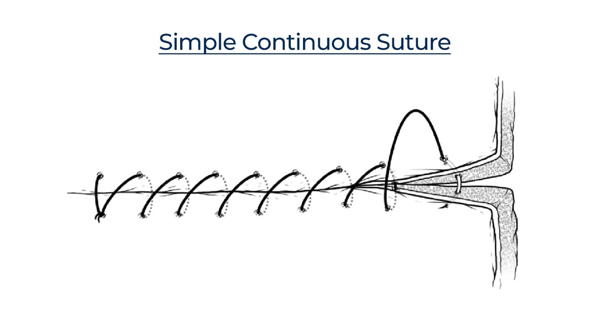
2. Simple Continuous:
A simple interrupted suture is placed as is done normally, and then the needle is reinserted in a continuous fashion such that the suture passes perpendicular to the incision line below and obliquely above. It is ended by passing a knot over the untightened end of the suture.
Advantages:
• Distributes tension uniformly across the entirety of the suture over the wound
• Only two knots are present in the suture: one at the beginning and one at the end, with associated tags
Disadvantages:
• If cut at one point, the suture thread loosens along the length of the wound, which then gapes open
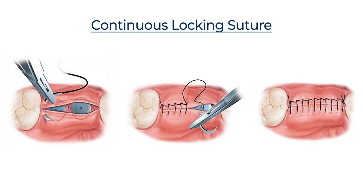
3. Continuous Locking / Blanket
The continuous locking or blanket suture technique is similar to the simple continuous type, but the locking is provided by withdrawing the suture through its own loop. It is indicated in long edentulous areas, the tuberosity, or retromolar areas.
Advantage:
• Reduced number of knots
• Uniform distribution of tension
• Prevents excessive tightening
Disadvantage:
• Prevents the adjustment of tension over the wound if tissue swelling occurs
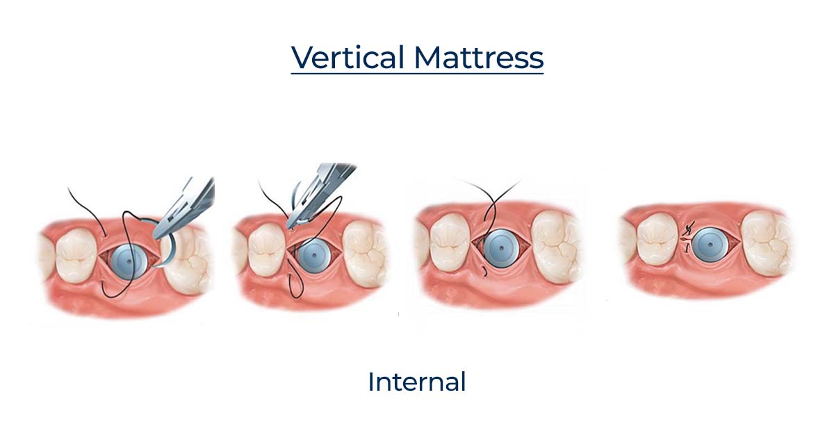
4. Vertical Mattress
a) Internal Vertical Mattress:
It passes at two levels — one deep, to provide support and approximation of wound surfaces at depth, and the other at a superficial level to draw the edges of the wound together and evert them. It is majorly used to close deep wounds.
Advantages:
• Better adaptation and maximum tissue approximation
• Slight eversion of wound margins
• In regions where wound healing may be delayed, it is better to give vertical sutures as they provide added support and help in controlling soft tissue hemorrhage
• Runs parallel to the blood supply of the edge of the flap and therefore doesn’t interfere with healing
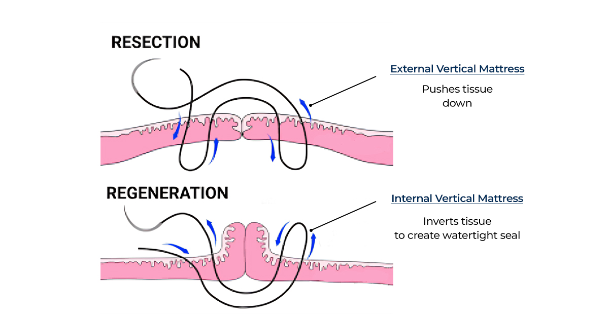
External Vertical Mattress:
The suture provides excellent wound support, promotes superior wound eversion, and decreases dead space. The needle is introduced 5–10 mm from the wound edge, and a deep bite of tissue is taken before exiting the flap on the opposite wound edge. The needle position is then reversed in the needle holder, and the needle is reintroduced 1–3 mm from the second side of the wound. A smaller bite of tissue is taken before exiting on the first side of the wound, and then a knot can be secured.
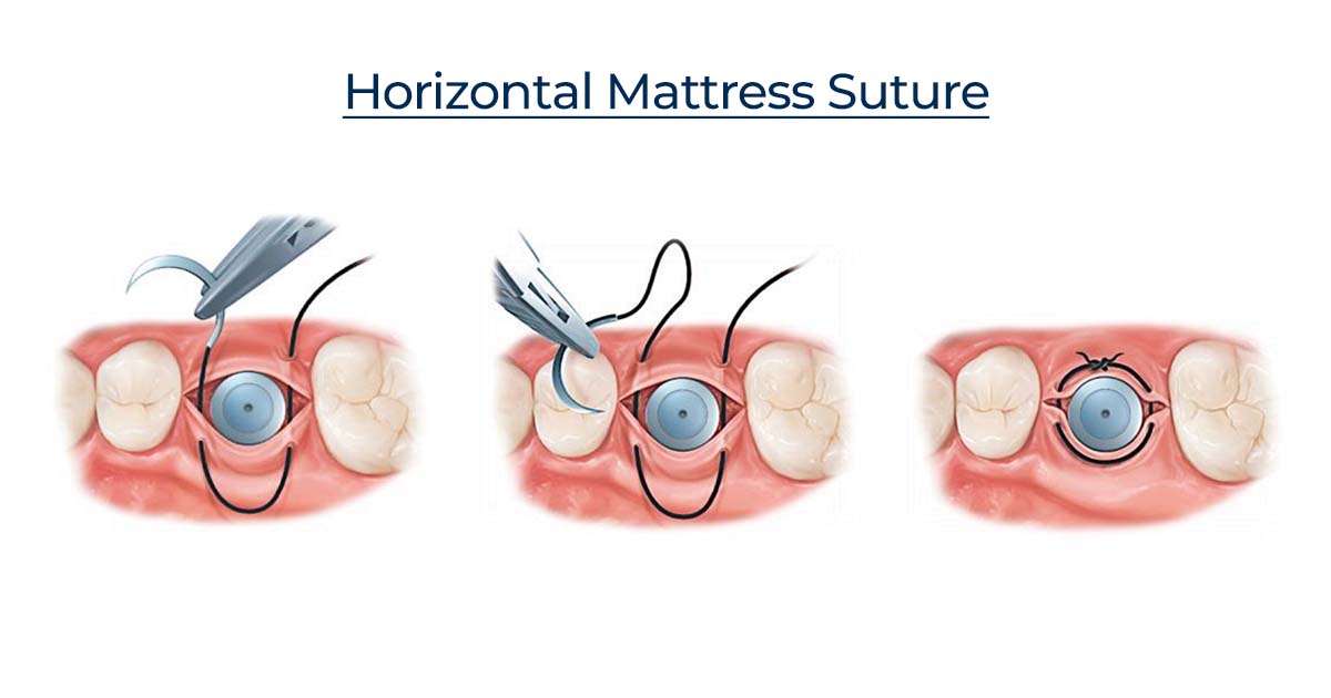
5. Horizontal Mattress
It helps in eversion of mucosal margins, bringing greater areas of raw tissue into contact. Its indicated use is for closing bony deficiencies such as oro-antral fistula or cystic cavities.
Advantages:
• Prevents the flap from being inverted into the cavity
• Helps control post-operative hemorrhage from the gingiva around the tooth socket by tensing the mucoperiosteum over the underlying bone
• Does not cut through the tissue, so can be used in cases where there is tissue tension (inadequate tissue)
Disadvantage:
• Constricts the blood supply to the edges of the incision, which can cause wound dehiscence and necrosis
• The needle is passed from one edge to the other and then again from the same side to the first one, and a knot is tied
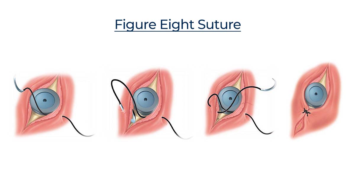
6. Figure of 8:
It is a modification of the horizontal mattress suture technique. While closing both sides of the soft tissue, this suture helps in the preservation of the position of the clot.
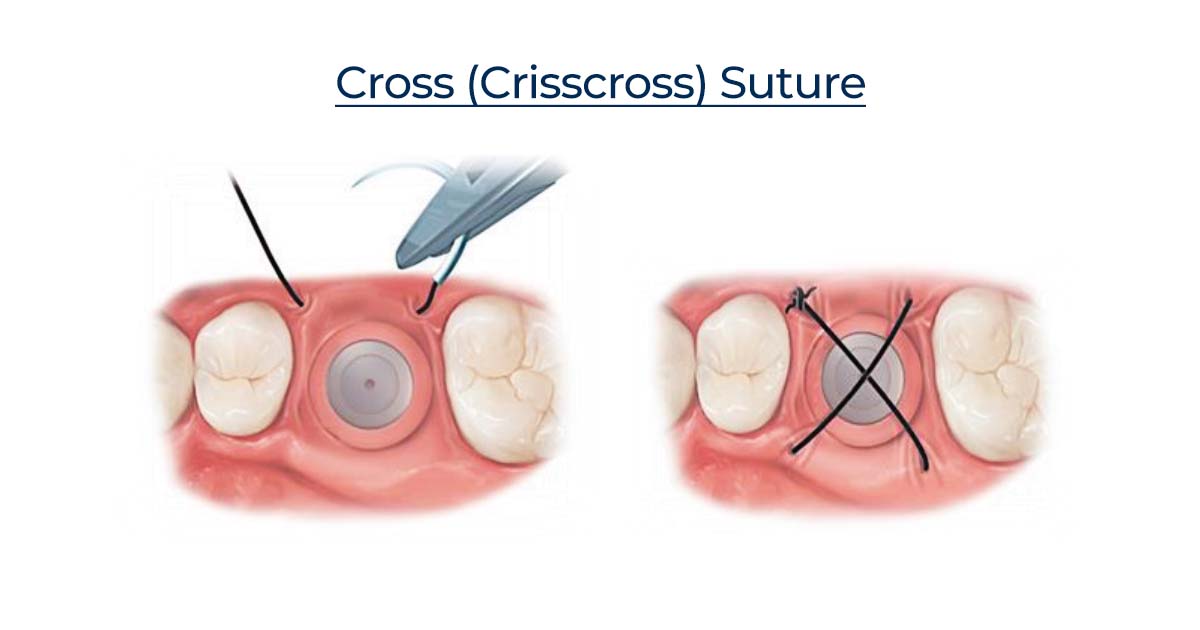
7. Crisscross:
This suture is given in a way that it forms a cross over the flap. A 3/8 needle is used to penetrate the tissue at the level of the mucogingival junction at the mesiobuccal line, travels horizontally under the flap, and emerges at the distobuccal line angle of the lingual aspect, such that the suture material crosses the surgical field and a knot is tied on the buccal aspect, forming a cross.
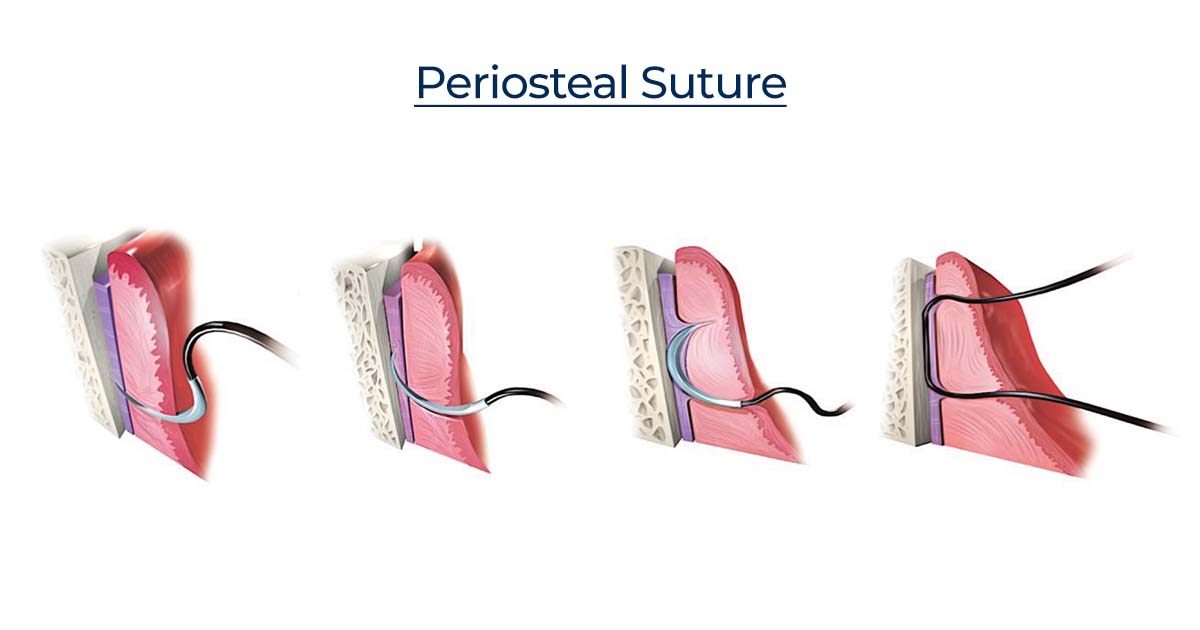
8. Periosteal Suturing Technique:
This technique involves the penetration of the needle into the periodontal/peri-implant tissues and periosteum all the way to the bone. This is followed by the rotation of the needle back to the direction it started, while penetrating through the periosteum again, then back through the keratinized tissue.
It is the 180-degree rotation of the needle grabbing the periosteum; the needle is moved along the bone below the periosteum, with rotation about the needle body, permitting the point to exit the periosteum and tissue.
Summary of Suturing Methods and Indications:
| S.No. | Suturing Methods | Indications |
| 1. | Simple interrupted | Commonly used; no specific indication |
| 2. | Simple Continuous | • Long wounds with minimal wound tension and good wound approximation • To secure split or full thickness grafts • Areas of cosmetic importance requiring less scarring. |
| 3. | Locking Continuous | Areas of moderate wound tension with good vascularisation but additional haemostasis e.g., scalp, post auricular, alveoloplasty. |
| 4. | Vertical Mattress | Areas that require wound eversion and to reduce wound tension eliminate dead space. |
| 5. | Horizontal Mattress | Provide strength and wound eversion and hence used in wounds under high tension. |
| 6. | Figure of 8 | Closure of extraction sites and other intraoral sites that require papillary adaption. |
| 7. | Crisscross | Stabilization of flaps over extraction sockets or grafted areas; provides cross-compression over the surgical field. |
| 8. | Periodontal | Stabilization and adaptation of periodontal or peri-implant soft tissue to underlying bone or implant surface. |
Conclusion:
Every thread and needle used in oral surgery contributes to the final outcome of healing, aesthetics, and patient comfort. From absorbable to non-absorbable options and simple interrupted to complex mattress techniques, the choice depends on the procedure, tissue type, and clinical scenario.
For dental professionals aiming to refine their surgical skills, understanding the characteristics of different sutures and when to use each technique can significantly elevate the quality of care. And when it comes to sourcing reliable and high-quality suturing materials, DentalKart serves as a one-stop destination. Whether you’re looking for suture threads, surgical needles, or complete suture kits, DentalKart offers a comprehensive range to support every oral surgical procedure.
After all, great closures make for great outcomes — and every well-placed suture brings your patient one step closer to a smooth, confident smile.
No Comment