Traditional Dentistry vs Modern Dentistry: The Challenges of Outdated Dental Education

Traditional Dentistry vs Modern Dentistry: The Challenges of Outdated Dental Education – Dentistry is a rapidly evolving field, with new materials, advanced equipment, and cutting-edge technologies being integrated every day. However, dental education in India continues to rely heavily on outdated methods, often requiring students to master older materials and techniques in the name of learning the basics. While understanding foundational concepts is important, this approach prevents students from exploring newer, more efficient technologies — the same ones they will eventually need when they step into clinical practice.
The gap between what is taught in dental schools and what is practiced in modern dentistry puts fresh graduates at a disadvantage. They are often forced to learn essential modern techniques on their own, either through expensive hands-on courses or trial and error in their private practice. This not only delays skill development but also affects patient care by limiting access to the best available treatments.
In this blog, we will highlight some outdated traditional methods still practiced in dental schools and explore modern alternatives that should be incorporated to bridge this gap and better equip future dentists.
Outdated Traditional Methods Still Practiced in Dental Schools:
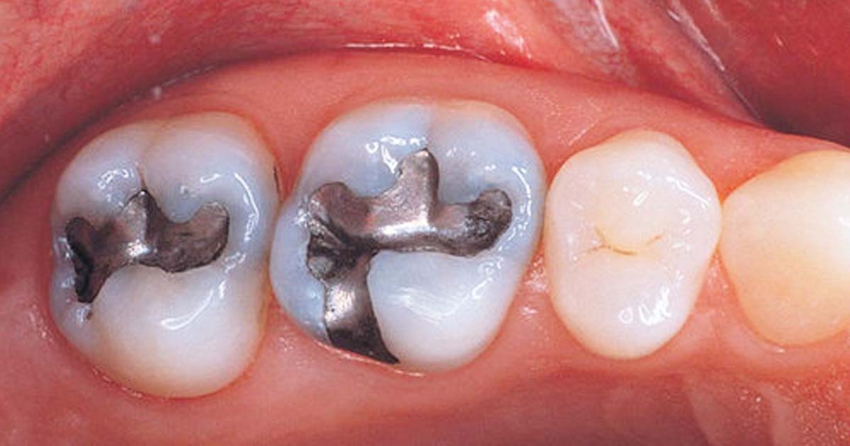
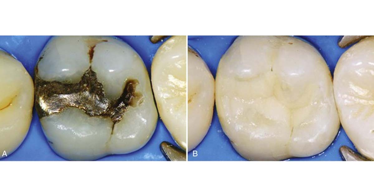
Amalgam restorations:
Amalgam restorations are taught in dental school right from the 2nd year of the dental course as pre-clinicals. From its manipulation to packing it in the cavity, everything is done by the students. This method is dangerous as the mercury present in the composition poses a threat not only to the student’s health but also to the dental assistants and teachers present during college hours.
Other than the amalgam usage during pre-clinicals, amalgam restorations are also performed during clinicals by these students when they are promoted to the final year. This increases the exposure to patients who will have mercury content in their oral cavity for years after the procedure is done in a dental college. Amalgam fillings not only weaken the tooth but are also capable of causing neurological impairment or kidney dysfunction.
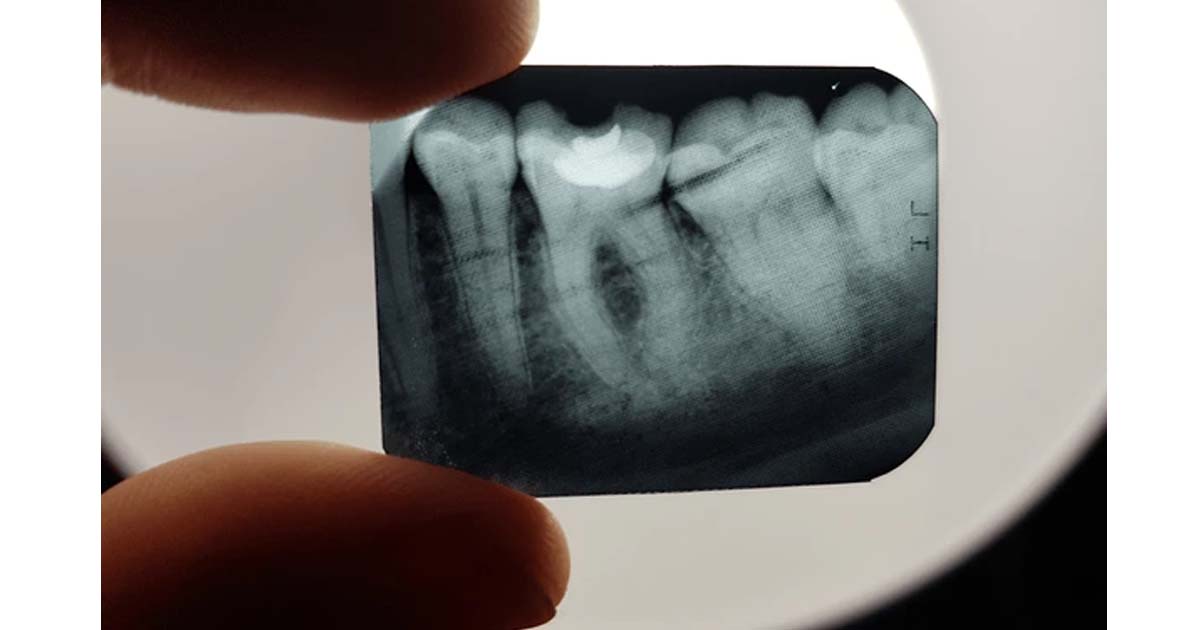
Traditional X-rays:
Traditional X-rays taught in colleges are of no use with the growing demands of digital dentistry in today’s world. Students are taught how to develop a dental film in separate developer and fixer solutions in dingy dark rooms. This not only increases the time of diagnosis but also wastes the precious time of students that could be utilized in learning other relevant clinical procedures. X-ray films also have a handling issue as they have to be kept safe until all of the patient’s sittings are completed. Other than that, there is a chance of confusion with different dental films for different patients.
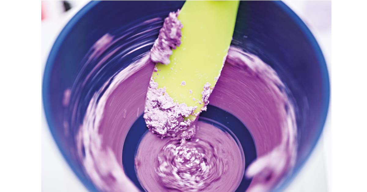
Alginate impressions for all cases:
Students are often forced to take only alginate impressions for all patients and for all types of cases requiring a positive replica of the patient’s teeth. Be it a partial denture patient or one requiring a crown, students are not taught the working of newer materials and equipment for better teeth impressions. These procedures lead to more material wastage during the learning years as students are not able to manipulate the materials as fast as experienced practitioners.
Other than material wastage, patients have a gag reflex, which can be quite a task for budding dentists to tackle, eventually decreasing their confidence. The time taken and the accuracy of these impressions are also not up to the mark many times. Furthermore, the shrinkage of the impression that occurs by the time it is poured leads to errors in the complete replication of the desired area.
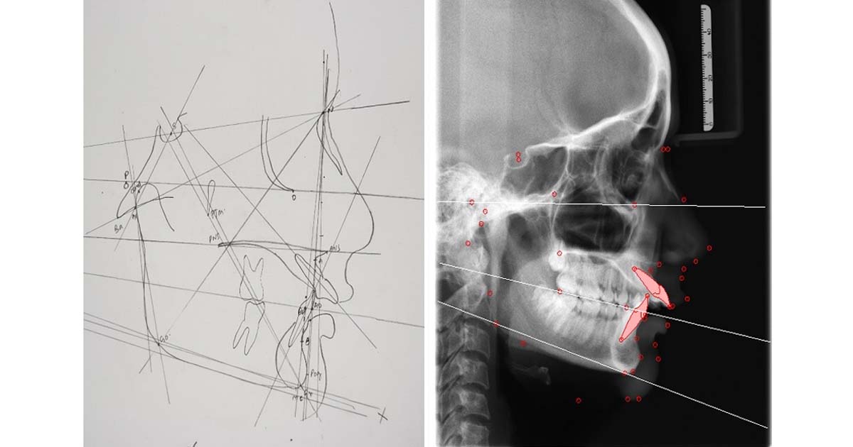
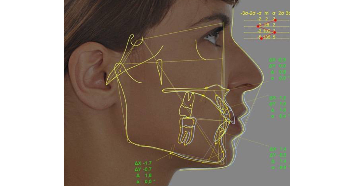
Cephalometric tracing by hand:
Not only is the manual tracing of cephalographs time-consuming, but it is also error-prone, as different persons interpret different points with varying degrees of precision due to inter-observer variability. The tracings are also done on delicate and thin sheets, which are often difficult to store and tear very easily.
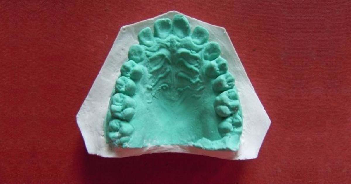
Traditional plaster casts:
The traditional plaster cast often has irregularities in texture and air bubbles. The replica lacks accuracy and doesn’t completely resemble the required area of the oral cavity to be worked on. Additionally, the trimming process increases dust particles in the air, leading to occupational hazards for those continuously working on them.
Modern Methods That Should Be Incorporated into Dental Education
New Restorative Materials:
With new materials being introduced frequently, it is easier for dentists to explore and learn new techniques. These materials are more biocompatible, safe, cost-effective, and easier to use than traditional restorative materials like amalgam. Restorative materials like Glass Ionomer Cement and resin-based composites, etc. can be used to restore dental cavities. These do not pose health hazards, as seen in the case of amalgam.
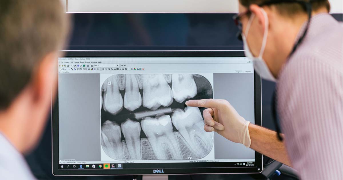
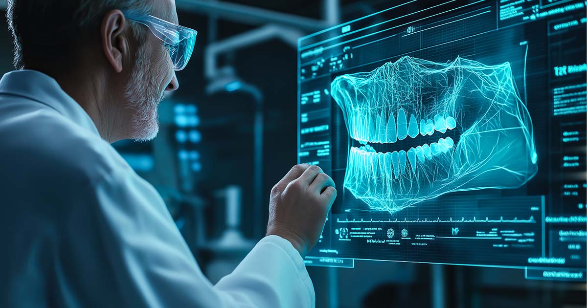
Digital Radiography:
It is a superior imaging technique used in dentistry, wherein electronic sensors generate images of the oral cavity. Students should be taught to handle this equipment regularly rather than relying on traditional films. The reduced processing time, superior image quality, and easy handling make it convenient for dental students and dentists to stay updated with newer technology. This ultimately helps in better diagnosis of dental diseases, leading to improved oral care.
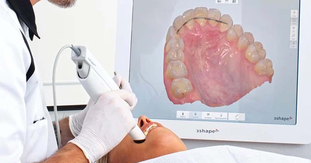
Integrating Digital Dentistry into the Curriculum
Integrating digital dentistry into the curriculum is essential for preparing future dentists to adopt modern dental practices. Dental colleges should incorporate digital workflows, such as computer-aided design and computer-aided manufacturing (CAD/CAM), intraoral scanners, and intraoral cameras, into their training programs. Students must learn to use digital scanners to capture highly accurate intraoral images, which can be converted into 3D models for designing and fabricating restorations like crowns and bridges with exceptional precision and efficiency.
Intraoral cameras further enhance diagnosis and treatment planning by providing high-resolution images of the oral cavity, improving patient communication and documentation. These technologies not only streamline workflows but also enhance patient satisfaction through superior detailing and high-quality restorations.
However, it is often not taught to dental students and is also not explored by many of them even when they start their dental clinic because of the high costs. If the dental colleges integrate this in their clinical syllabus and make it available for their students right in the beginning they can prioritize this as a need for their dental practices and buy it first whenever setting up their practice.
Artificial Intelligence in Dental Diagnosis
Artificial intelligence (AI) is transforming dental diagnosis by enabling precise analysis of radiographic images, early anomaly detection, and improved treatment planning. By leveraging machine learning, dentists can enhance diagnostic accuracy and develop personalized treatment plans, ultimately improving patient outcomes.
Read Also: Artificial Intelligence in Dentistry: A Glimpse into the Future
The integration of advanced technology, such as AI and 3D printing, has revolutionized modern dentistry, with research playing a key role in these advancements. From AI-driven diagnostics to stronger, more durable restorative materials like composites and ceramics, ongoing research enhances the aesthetics and longevity of treatments. By incorporating these innovations into dental education, future professionals can become proficient in newer equipment and techniques, ensuring better patient care and long-lasting results.
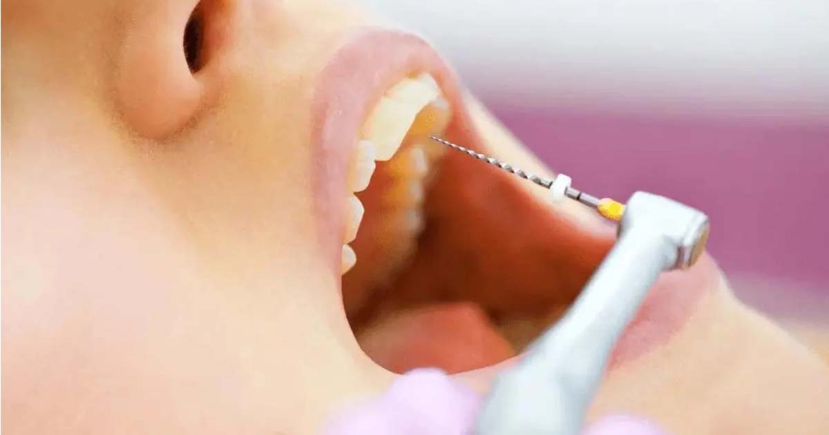
Endomotor and Single-Sitting RCTs
Often taught theoretically but never practically, endomotors and their use in single-sitting RCTs are left for graduates to explore only after leaving college. They are often compelled to learn these skills through hands-on courses from senior dentists, leading to additional learning costs. These techniques are glorified in colleges as postgraduate-level skills, but when B.D.S. graduates enter the dental market, they are often criticized for not knowing them, leading to demotivation due to a lack of skills and ultimately compromising on salary expectations.
New Software for Cephalometric Tracing
This new technology is not given enough importance, but cephalometric tracing of digital radiographs and photographs through software is one of the major advancements in orthodontic treatment. It helps assess a patient’s growth during orthodontic procedures. Not only does it save time and storage space, as compared to manual tracing on cephalometric films, but it also improves analysis accuracy by providing precise soft and hard tissue markings. There are minimal chances of inter-observer variability, ensuring consistent and accurate results with every cephalometric analysis.
The Role of Dental Colleges in Driving Innovation
With over 400 dental colleges in India, the demand for dental services is increasing daily, along with greater oral health awareness and the rise of dental tourism. Despite these advancements, new technologies still face challenges in being adopted and taught in dental colleges.
One of the primary obstacles is the high cost of implementation, both during dental education and clinical practice. Many of these technologies are imported, making them financially inaccessible for many institutions. Additionally, the lack of proper guidance and training hinders their effective use. Even though these technologies are designed to be safe and user-friendly, improper training can lead to inefficient or even harmful practices.
To overcome these barriers, it is essential to integrate these advancements into the dental curriculum. By doing so, future dentists can explore, understand, and recognize the necessity of these innovations. Modern technology not only saves time in diagnosis and treatment but also enhances learning through engaging visuals and interactive devices. This, in turn, helps students develop their skills effectively, ensuring they remain updated with the latest advancements in the Indian dental fraternity.
No Comment